Meet the 2021 Knight Campus Undergraduate Scholars
The Knight Campus Undergraduate Scholars Program pairs promising undergraduates with research mentors — graduate students, postdocs, and faculty members — immersing them in a 12-month, comprehensive research experience in Knight Campus-affiliated labs.
While these young scientists are taking on independent research projects in a diverse set of fields, they are also finding creative ways to conduct their research remotely. Below are some examples of what the cohort has been working on this term.
Maybelle Clark Macdonald Scholar
Hometown: Beaverton, Oregon
Class: Sophomore
Major: Human Physiology
Mentor: Parisa Hosseinzadeh, Assistant Professor at the Knight Campus
Lab: Hosseinzadeh Lab
Designing Peptide Binders for the Detection of Periodontal Disease
“With guidance from my mentor Parisa Hosseinzadeh, I've been working on designing peptide binders that will accurately and selectively bind to the protein MMP-8. I've used computational design methods using software such as Rosetta and PyMol, which allow me to generate and visualize designs. The image shown here is a heat map that display the effects of various mutations to my peptide binder. I used heat maps like this to analyze which mutations are the most energetically favorable while also maximizing the shape complementarity at the protein-binder interface.
As a Knight Campus Undergraduate Scholar, my goal is to use the results of my research to improve the prognosis of periodontal disease in medical patients. The ultimate goal of this project is to incorporate my peptide binder design as a detection module in biosensors. Salivary MMP-8 has been observed as a biomarker in medical patients with periodontal disease. By designing a binder that can effectively bind to MMP-8, there will be an effective way to detect periodontal disease in patients who either have periodontal disease or are susceptible to periodontal disease.
With the help of my mentor Parisa Hosseinzadeh, I’ve learned both lab skills as well as computational skills that will prepare me for my career in medical research. I leave the lab every single day feeling inspired and with a profound sense of purpose.”


Maybelle Clark Macdonald Scholar
Hometown: Portland, Oregon
Class: Junior
Major: Biochemistry
Mentor: Julia Fehr, Graduate Student in the Jasti Lab
Lab: Jasti Lab
Identifying Potential Imaging Reagents to Detect Diseases and Disorders
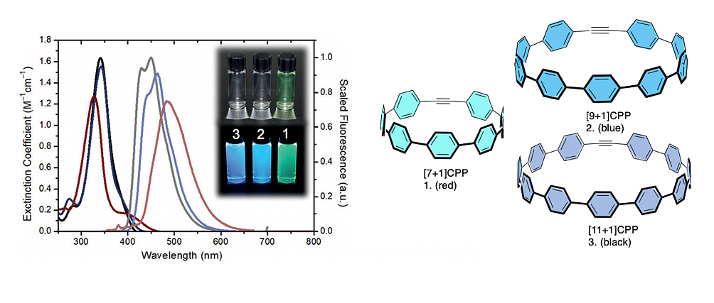
“This is a screenshot of a newly synthesized alkyne-containing cycloparaphenylene ([n+1]CPP). These organic molecules have unique size-dependent optical and electronic properties. My research as a Knight Campus Undergraduate Scholar focuses on their potential to be utilized as bioorthogonal fluorophores also known as labelling reagents.
Over the next year, I will be performing various proof of concept biology reactions including labeling a biomolecule with a [n+1]CPP. If I am able to attach these [n+1]CPPs to specific biomolecules, it will be a huge step in determining their potential as bioorthogonal imaging reagents capable of detecting many diseases and disorders.”


Guldberg Scholar
Hometown: Portland, Oregon
Class: Junior
Major: Psychology
Mentor: Beth Bearce, Postdoctoral Scholar in the Grimes Lab
Lab: Grimes Lab
Discovering how Motile Cilia Regulates the Body Shape of Zebrafish
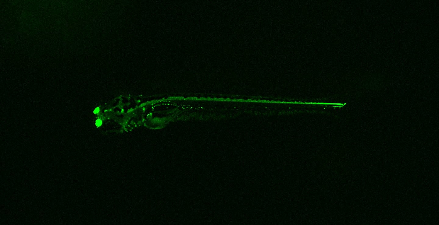
“This is an image of a two-day old zebrafish that expresses a green fluorescent protein in cells that have motile cilia. Motile cilia are microtubule-based, hair-like organelles that protrude from the cell surface and beat in a wave-like motion to generate fluid flows. These flows are critical for the function of many organ systems in both fish and humans, including the central nervous system and kidneys. This transgenic fish shows bright green fluorescence in the nose, brain, early spinal column, and pronephros (early kidney-like structures).
My mentor and colleagues recently showed that cerebral spinal fluid flow, driven by motile cilia in the brain ventricles and spinal canal, is a critical regulator of body-axis shape in zebrafish. I am interested in exploring the pathways that occur downstream of these central nervous system motile cilia, to ultimately allow the embryo to sense, create, and maintain the correct body shape.”
Petrone Scholar
Hometown: Eugene, Oregon
Class: Junior
Major: Chemistry and Sociology
Mentor: Thaís de Faria, Graduate Student in the Johnson and Haley Lab
Labs: Johnson Lab and Haley Lab
Studying the Rates of Reactive Hydrosulfide with the Assistance of a Supramolecular Receptor
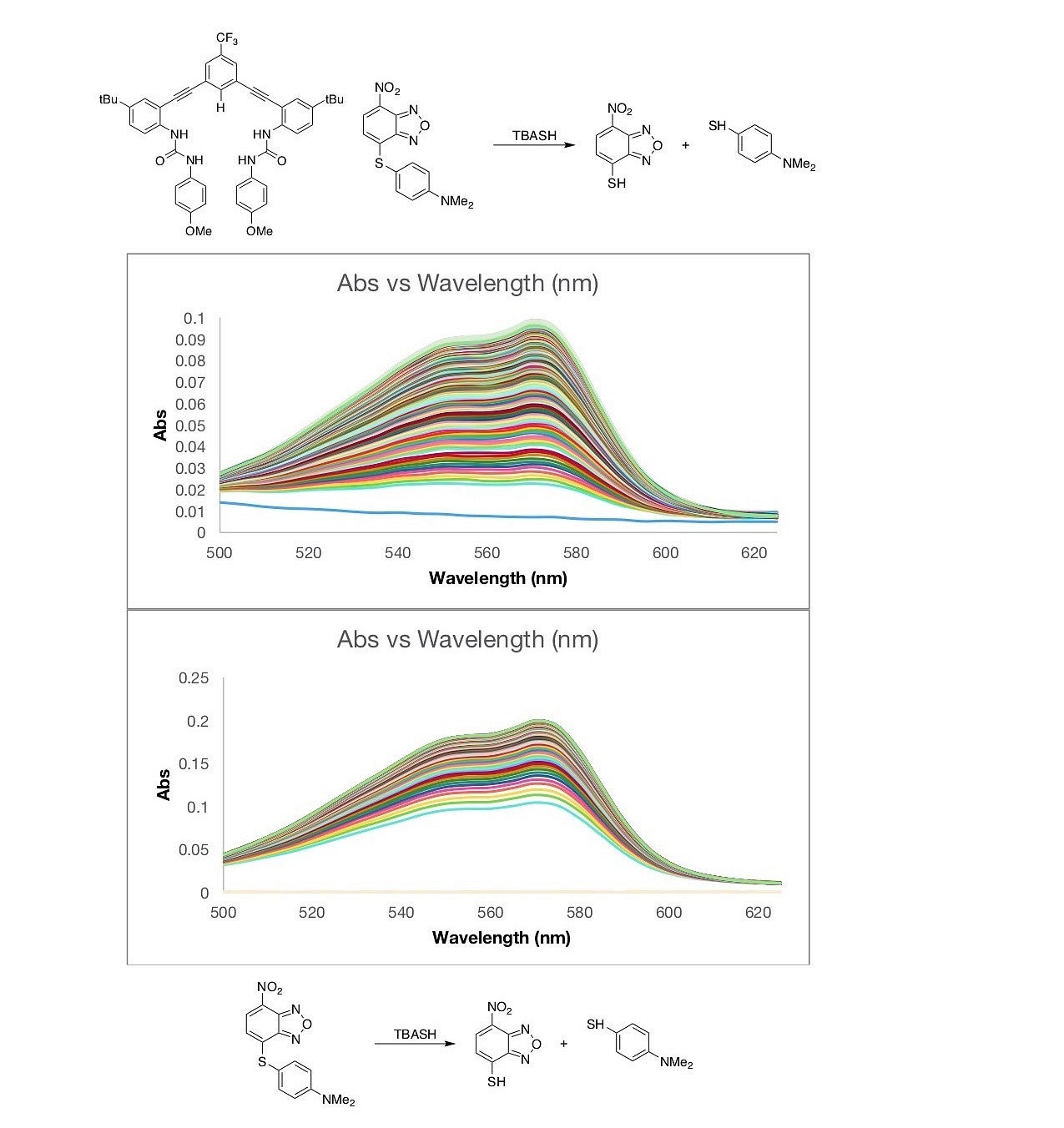
“As a Knight Campus Undergraduate Scholar, I am researching the kinetics of a nitrobenzofurazan (NBD) thioether undergoing a nucleophilic aromatic substitution (SNAr) reaction with the addition of the highly reactive anion HS-. I am studying how the kinetics differ with and without the addition of a supramolecular receptor to see how non-covalent interactions affect the rate of the reaction. This study can provide insight into how our bodies might stabilize HS- through non-covalent interactions and give us a better understanding of the behavior of this species in biological systems.
The bottom image seen here is a UV-Vis spectrum of the NBD-thioether after the immediate injection of HS-, using TBASH as a source of the anion. The growth seen in the spectrum shows the reaction proceeding. Once it reaches a peak, we know the reaction has gone to completion. The top image shows the same reaction but with the addition of the supramolecular receptor. It can visibly be seen that the reaction takes longer to reach completion when the receptor is in solution, as there are more lines proceeding the peak. I take this data and create non-linearized plots to obtain the quantitive rate kinetics to see how the receptor affects the speed of the reaction.”
Maybelle Clark Macdonald Scholar
Hometown: Salem, Oregon
Class: Junior
Major: Chemistry
Mentor: Jeremy Bard, Graduate Student in the Haley Lab
Lab: Haley Lab
Synthesizing and Characterizing Molecules to Localize Cell Lysosome
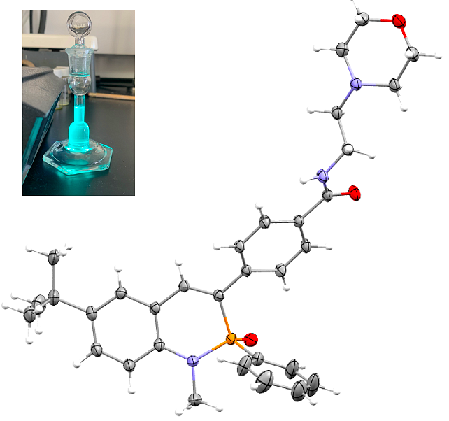
“In the Haley Lab, I am working on the PN heterocycle project, where we are synthesizing and studying phosphorous nitrogen-containing fluorescent dyes. Our current objective is to take one of these fluorescent molecules and modify its structure so it can be put inside of cells. The morpholine targeting group we have attached to the molecular structure will allow it to localize to the cell's lysosome. Once it is inside the lysosome of the cell, the fluorescent properties of the molecule lets researchers monitor cellular processes as they happen.
Collecting this kind of real time information is extremely important for molecular biologists who are studying diseases caused by cellular dysfunction. My mentor Jeremy and I are handling the synthesis and characterization of our target molecule, while our collaborators in the Pluth lab are using what we have made to actually image cells. This image shows the structure of our target molecule, which we recently collected using x-ray diffraction. The morpholine targeting group is on the upper right side of the molecule, which will is responsible for guiding the molecule to the lysosome. The glowing vial in the inset image is what the molecule looks like when it is fluorescing.”
Maybelle Clark Macdonald Scholar
Hometown: Portland, OR
Class: Junior
Major: Biology
Mentor: Marian Hettiaratchi, Assistant Professor at the Knight Campus
Lab: Hettiaratchi Lab
Developing Hydrogels for Bone Regeneration
“The Hettiaratchi Lab aims to combine chemical and biomedical engineering to design biomaterials that control protein delivery to injured tissues. My current project is focusing on designing and testing the cytocompatibility of various types of hydrogel biomaterials composed of natural polymers, like collagen or hyaluronic acid, that can ultimately be used to deliver osteogenic proteins for bone regeneration. In the future, expansions of this project could lead to the use of hydrogels as a biomaterial that rivals the healing response of bone autografts (tissue grafts from the same individual).
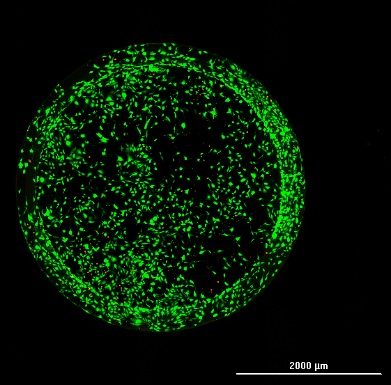
This is an image of oxidized and carbohydrazide hyaluronic acid hydrogels formed by dynamic, covalent bonds seeded with 3T3 fibroblast cells five days post-treatment. Live cells were evaluated using green fluorescence from GFP and dead cells were evaluated using red fluorescence from ethidium homodimer. This particular hydrogel showed high cell viability and a rate of cell growth that surpassed all other conditions.”


Knight Scholar
Hometown: Anchorage, Alaska
Class: Junior
Major: Human Physiology and Spanish
Mentor: Mitchell Fisher, Graduate Student in the Greenhouse Lab
Lab: Greenhouse Lab (Action Control Lab)
Identifying the Role of a Global Stopping Mechanism in the Partial Cancellation of Unitary Movements
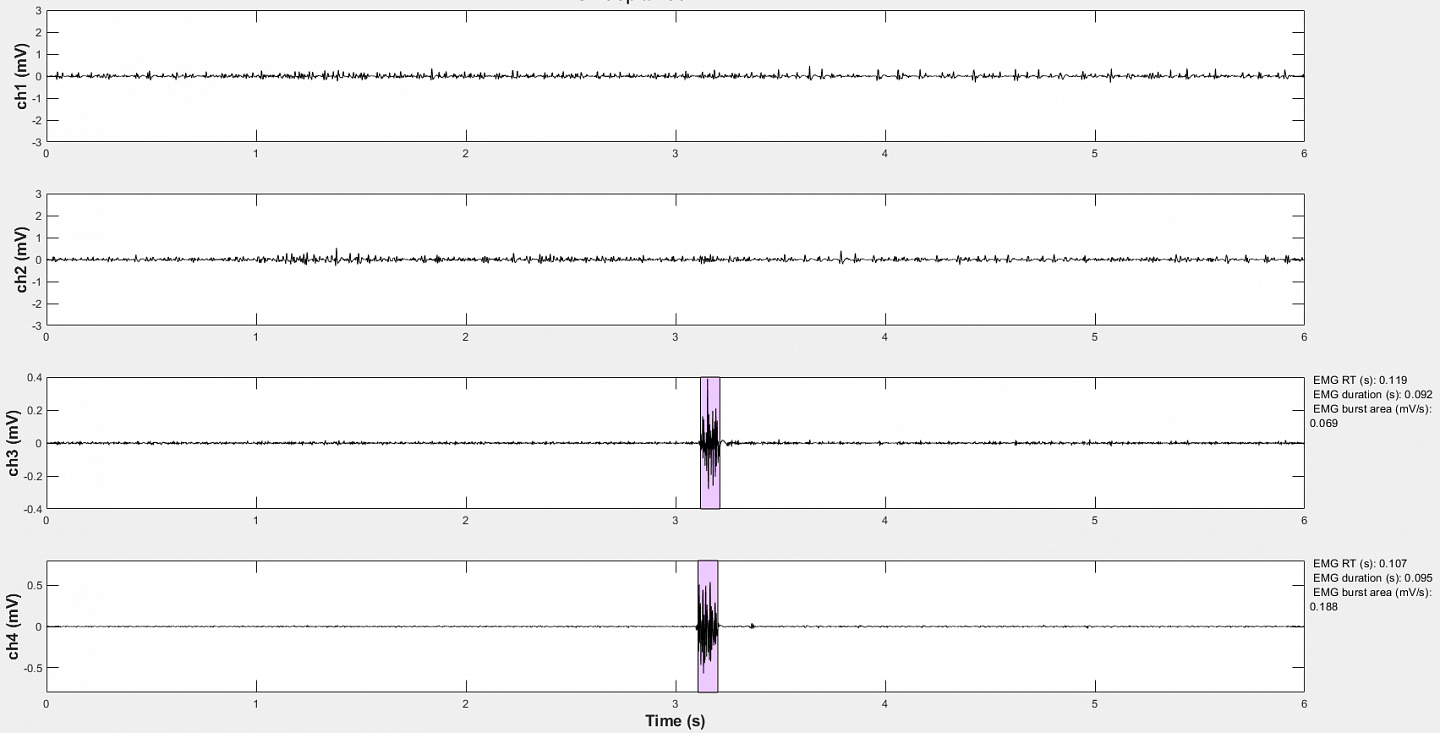
“My research in the Greenhouse Lab is geared towards investigating how the brain controls human movement. In our current project, we are investigating movement cancellation and how cancelling one action can affect movements that are being performed concurrently. This image shows surface electromyography (EMG) data, a measure of electrical activity in the muscle, from one trial of our project’s pilot data collection. The two highlighted bursts show the subject activating both their left and right first dorsal interosseous muscles to lift their index fingers at the three second mark. When we collect data in the future, the first and second channels will be recording EMG activity produced by constant contraction of the left and right abductor digiti minimi, a muscle that controls the little finger. A fraction of the trials will incorporate a signal prompting subjects to cancel the index finger lift just before they execute it.
The end goal of this study is to compare the amplitude of the abductor digiti minimi EMG output between cancellation and go trials to look for evidence of partial action cancellation leading to global inhibition of muscle activity.”
Maybelle Clark Macdonald Scholar
Hometown: Beaverton, Oregon
Class: First-year
Major: Biochemistry
Mentor: Hiro Uehara, Senior Research Associate in the Ambati Lab
Lab: Ambati Lab
Investigating the Structure of the Guttae in Corneas to Develop Medical Treatments and Preventative Measures
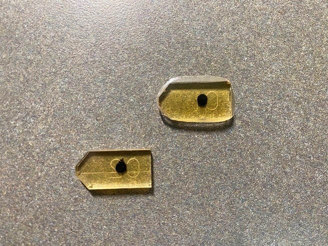
“In the Ambati Lab, I am researching the unique structure of the endothelial layer of corneas with Fuchs’ Endothelial Corneal Dystrophy (FECD) through the use of a Focused Ion-Beam and Scanning Electron Microscopy (FIB-SEM). FECD is when the cornea fills up with fluid by damaging the endothelial layer which leads to corneal edema, opacity and vision loss. But very little is known about precise structural aspects of FECD, especially guttae which are excrescences of corneal collagen. To study this, a plasma-Focused Ion Beam microscope (or plasma-FIB microscope) will take micro-cross sections of our sample using an ion source and develop high-definition images.
This is an image of a cornea that has been stained with uranyl acetate and osmium tetroxide and embedded in epoxy resin so that we can obtain images by the FIB-SEM. The staining process creates a contrast between the cornea and any extra tissue so that our target area is highlighted for the microscope. Later this term, I will be learning how to use a FIB-SEM so that I can successfully image the guttae structure in the corneal endothelium and develop a plan of action based on the results.
This research would give the medical community a better understanding of the structural aspects of corneas with FECD and help understand disease developing mechanisms and preventative measures. I am grateful for the valuable experience I am gaining through the opportunity to work with my mentor Hiro Uehara on this project and from the support of the other researchers in the Ambati Lab.”

Clark Honors College Scholar
Hometown:Portland, Oregon
Class: Junior
Major: Biology
Mentor: David Garcia, Assistant Professor in the Department of Biology
Lab: Garcia Lab
Identifying Prion Proteins as a Potentially Beneficial Epigenetic Mechanism
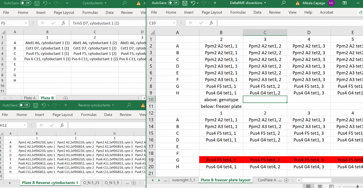
“My research in the Garcia Lab aims to confirm the identity of novel prion proteins in the yeast proteome. To do this, I trace the inheritance patterns of the traits caused by the putative prion proteins I study. The first step of this process, which I am currently working on, involves crossing strains of yeast, isolating the offspring I want to study, and freezing the strains into stocks for future experiments.
This is an image of the Excel files where I keep track of the strains. Next, I will be determining how the traits inherited in the yeast by performing growth assays. The goal of my research is to ultimately identify prion proteins as a potentially beneficial epigenetic mechanism that would allow cells to respond to special conditions in their environment.”

Knight Scholar
Hometown: Damascus, Oregon
Class: Junior
Major: Biology
Mentor: Nicole Kurhanewicz, Postdoctoral Scholar in the Libuda Lab
Lab: Libuda Lab
Characterizing the Impacts of Heat Shock Factors on Genomic Integrity
“I am studying the mechanisms behind how heat stress produces DNA damage in developing sex cells. As part of the Knight Campus Undergraduate Scholars program, I am investigating the potential involvement of the Heat Shock Response pathway, and its master regulator HSF-1, which seems to provide oocyte-specific protection against heat-induced DNA damage.
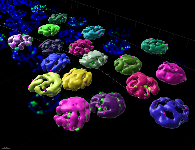
To quantify DNA damage, I use immunofluorescence microscopy. In this 3D-image of a male germline, casts of individual sperm nuclei are rendered in different colors using image analysis software, and the DNA damage is marked by small pink or green spots. Using this software, I can count the number of sites of DNA damage within a single nucleus. My research employs this high-resolution microscopy approach in combination with transcriptomic analysis to identify candidate proteins and pathways that respond differently to heat in developing sperm and eggs.
After spending another year with the Libuda Lab, I will pursue a Ph.D., and eventually hope to start my own molecular biology lab.”
Clark Honors College Scholar
Hometown: Portland, Oregon
Class: Junior
Major: Biology
Mentor: Zoe Irons, Graduate Student in the Grimes Lab
Lab: Grimes Lab
Understanding the Dynamics of Body Straightening and Developmental Growth
“In the Grimes Lab, I am learning how we form the right shape during development and growth. Development involves the precise positioning of cells at the right time and place to form a functional organism.
Through my work as a Knight Campus Undergraduate Scholar, I am investigating the dynamics of body straightening through zebrafish. This image shows several mutants defective for a gene necessary for bodily straightening (cfap298). These mutants develop phenotypes similar to adult idiopathic scoliosis in humans. I will be taking time-lapse videos of how these mutants develop in order to better understand how the body straightening process naturally happens, and how that process can mechanistically go wrong to produce a disease such as idiopathic scoliosis.”
Petrone Scholar
Hometown: Beaverton, OR
Class: Sophomore
Major: Biology
Mentor: Osamu Iwasaki, Research Assistant Professor in the Noma Lab
Lab: Noma Lab
Identifying 3D Genome Structures to Determine the Function of Condensin in Chromosomal Segregation
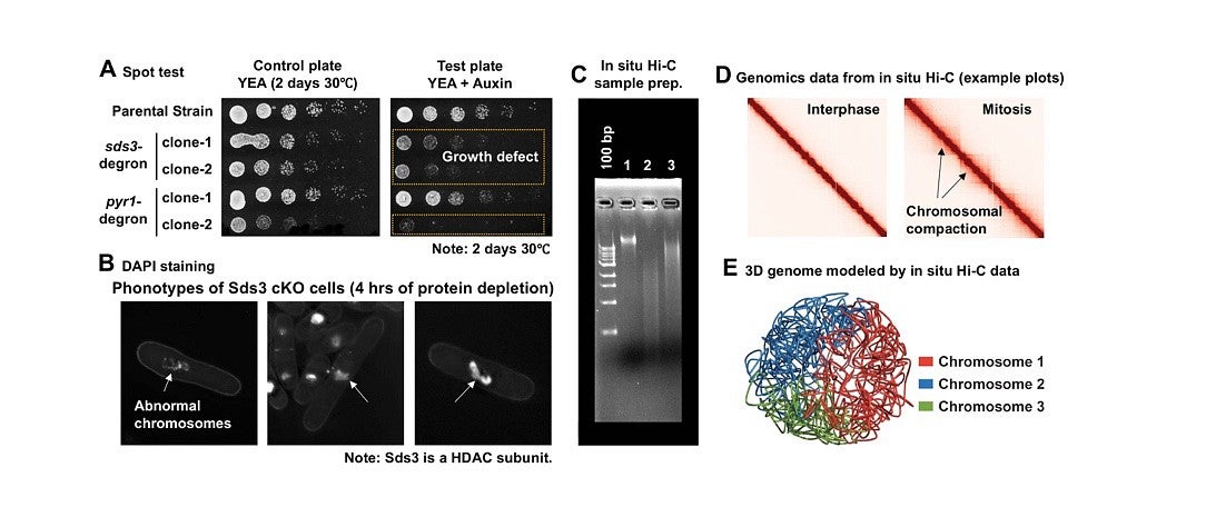
“In the Noma Lab, I am studying how the 3D genome structure is organized by condensin and its interactors using the fission yeast model organism. The condensin protein complex is known as a key genome organizer and functions in chromosomal segregation during mitosis. My project is to determine how condensin interactors, such as Sds3 histone deacetylase and Pyr1 pyruvate carboxylase, are involved in 3D genome organization. To do this, I have constructed auxin-inducible degron strains that conditionally knock out proteins of interest. In this image, the spot tests (A) show significant growth defects of knockout strains in the presence of auxin, indicating that the essential proteins have been successfully knocked out by the degron system. The microscope images (B) show the effects of the conditional knock-out (cKO) of the Sds3 protein on chromosomal segregation during mitosis. This data potentially suggests that Sds3 interacts with condensin to form the specific 3D genome structure that is required for the fidelity of chromosomal segregation. After these knockout strains were constructed, I performed in situ Hi-C to determine the effect of the depletion of condensin interactors on genome organization. The gel image (C) shows this process with the products after cross-linking (1), restriction digestion (2), and ligation (3). My in situ Hi-C samples will soon be analyzed by a next-generation sequencer at the UO’s Genomics and Cell Characterization Core Facility. I am looking forward to seeing the resulting genomic data which will represent the structure of the entire fission yeast genome. (Examples of in situ Hi-C data reflecting the interphase and mitotic genome structures are shown (D).)
Using this data, I will be able to model the 3D genome structure (E). Repeating these procedures with other proteins of interest will allow me to determine how condensin interactors function in the organization of the 3D genome structure required for proper chromosomal segregation. I am thankful to be a part of this project because 3D genome organization is an exciting new area of research from which many fundamental discoveries are expected to emerge.”
